Anterior segment of eyeball
Front third of the eye
| Anterior segment of eyeball | |
|---|---|
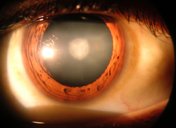 Human eye Anterior Segment - Magnified view seen on examination with a slit lamp under diffuse illumination showing conjunctiva overlying the white sclera, transparent cornea, pharmacologically dilated pupil and cataract | |
| Details | |
| Identifiers | |
| Latin | segmentum anterius bulbi oculi |
| MeSH | D000869 |
| Anatomical terminology [edit on Wikidata] | |

1. Lens, 2. Zonule of Zinn or ciliary zonule, 3. Posterior chamber and 4. Anterior chamber with 5. Aqueous humour flow; 6. Pupil, 7. Corneosclera with 8. Cornea, 9. Trabecular meshwork and Schlemm's canal. 10. Corneal limbus and 11. Sclera; 12. Conjunctiva, 13. Uvea with 14. Iris, 15. Ciliary body.
The anterior segment or anterior cavity[1] is the front third of the eye that includes the structures in front of the vitreous humour: the cornea, iris, ciliary body, and lens.[2][3]
Within the anterior segment are two fluid-filled spaces:
- the anterior chamber between the posterior surface of the cornea (i.e. the corneal endothelium) and the iris.
- the posterior chamber between the iris and the front face of the vitreous.[2]
Aqueous humour fills these spaces within the anterior segment and provides nutrients to the surrounding structures.
Some ophthalmologists and optometrists specialize in the treatment and management of anterior segment disorders and diseases.[3]
See also
References
- ^ "WikiJournal of Medicine/Medical gallery of Blausen Medical 2014 - Wikiversity".
- ^ a b Cassin, B. and Solomon, S. Dictionary of Eye Terminology. Gainesville, Florida: Triad Publishing Company, 1990.
- ^ a b "Departments. Anterior segment." Archived September 27, 2006, at the Wayback Machine Cantabrian Institute of Ophthalmology.
External links
- Anterior+Eye+Segment at the U.S. National Library of Medicine Medical Subject Headings (MeSH)
- v
- t
- e
Anatomy of the globe of the human eye
(outer)
| Sclera | |
|---|---|
| Cornea |

tunic (middle)
| Choroid | |
|---|---|
| Ciliary body | |
| Iris |
| Layers | |
|---|---|
| Cells |
|
| Other |
of the eye
| Anterior segment | |
|---|---|
| Posterior segment |
- Keratocytes
- Ocular immune system
- Optical coherence tomography
- Eye care professional
- Eye disease
- Refractive error
- Accommodation
- Physiological Optics
- Visual perception
| Authority control databases: National |
|
|---|
 | This article about the eye is a stub. You can help Wikipedia by expanding it. |
- v
- t
- e


