BAIAP2
Protein-coding gene in the species Homo sapiens
| BAIAP2 | |||||||||||||||||||||||||||||||||||||||||||||||||||
|---|---|---|---|---|---|---|---|---|---|---|---|---|---|---|---|---|---|---|---|---|---|---|---|---|---|---|---|---|---|---|---|---|---|---|---|---|---|---|---|---|---|---|---|---|---|---|---|---|---|---|---|
 | |||||||||||||||||||||||||||||||||||||||||||||||||||
| Identifiers | |||||||||||||||||||||||||||||||||||||||||||||||||||
| Aliases | BAIAP2, BAP2, FLAF3, IRSP53, BAI1 associated protein 2, BAR/IMD domain containing adaptor protein 2, WAML | ||||||||||||||||||||||||||||||||||||||||||||||||||
| External IDs | OMIM: 605475; MGI: 2137336; HomoloGene: 9697; GeneCards: BAIAP2; OMA:BAIAP2 - orthologs | ||||||||||||||||||||||||||||||||||||||||||||||||||
| |||||||||||||||||||||||||||||||||||||||||||||||||||
| |||||||||||||||||||||||||||||||||||||||||||||||||||
| |||||||||||||||||||||||||||||||||||||||||||||||||||
| |||||||||||||||||||||||||||||||||||||||||||||||||||
| |||||||||||||||||||||||||||||||||||||||||||||||||||
| Wikidata | |||||||||||||||||||||||||||||||||||||||||||||||||||
| |||||||||||||||||||||||||||||||||||||||||||||||||||


Brain-specific angiogenesis inhibitor 1-associated protein 2 is a protein that in humans is encoded by the BAIAP2 gene.[5][6]
Function
The protein encoded by this gene has been identified as a brain-specific angiogenesis inhibitor (BAI1)-binding protein. This interaction at the cytoplasmic membrane is crucial to the function of this protein, which may be involved in neuronal growth-cone guidance. This protein functions as an insulin receptor tyrosine kinase substrate and suggests a role for insulin in the central nervous system. This protein has also been identified as interacting with the dentatorubral-pallidoluysian atrophy gene, which is associated with an autosomal dominant neurodegenerative disease. It also associates with a downstream effector of Rho small G proteins, which is associated with the formation of stress fibers and cytokinesis. Alternative splicing of the 3'-end of this gene results in three products of undetermined function.[6]
Interactions
BAIAP2 has been shown to interact with:
References
- ^ a b c GRCh38: Ensembl release 89: ENSG00000175866 – Ensembl, May 2017
- ^ a b c GRCm38: Ensembl release 89: ENSMUSG00000025372 – Ensembl, May 2017
- ^ "Human PubMed Reference:". National Center for Biotechnology Information, U.S. National Library of Medicine.
- ^ "Mouse PubMed Reference:". National Center for Biotechnology Information, U.S. National Library of Medicine.
- ^ Oda K, Shiratsuchi T, Nishimori H, Inazawa J, Yoshikawa H, Taketani Y, Nakamura Y, Tokino T (June 1999). "Identification of BAIAP2 (BAI-associated protein 2), a novel human homologue of hamster IRSp53, whose SH3 domain interacts with the cytoplasmic domain of BAI1". Cytogenet Cell Genet. 84 (1–2): 75–82. doi:10.1159/000015219. PMID 10343108. S2CID 27688560.
- ^ a b "Entrez Gene: BAIAP2 BAI1-associated protein 2".
- ^ Okamura-Oho Y, Miyashita T, Ohmi K, Yamada M (June 1999). "Dentatorubral-pallidoluysian atrophy protein interacts through a proline-rich region near polyglutamine with the SH3 domain of an insulin receptor tyrosine kinase substrate". Hum. Mol. Genet. 8 (6): 947–57. doi:10.1093/hmg/8.6.947. PMID 10332026.
- ^ a b c d Miki H, Yamaguchi H, Suetsugu S, Takenawa T (Dec 2000). "IRSp53 is an essential intermediate between Rac and WAVE in the regulation of membrane ruffling". Nature. 408 (6813): 732–5. Bibcode:2000Natur.408..732M. doi:10.1038/35047107. PMID 11130076. S2CID 4426046.
- ^ a b Soltau M, Richter D, Kreienkamp HJ (Dec 2002). "The insulin receptor substrate IRSp53 links postsynaptic shank1 to the small G-protein cdc42". Mol. Cell. Neurosci. 21 (4): 575–83. doi:10.1006/mcne.2002.1201. PMID 12504591. S2CID 572407.
- ^ Krugmann S, Jordens I, Gevaert K, Driessens M, Vandekerckhove J, Hall A (October 2001). "Cdc42 induces filopodia by promoting the formation of an IRSp53:Mena complex". Curr. Biol. 11 (21): 1645–55. doi:10.1016/S0960-9822(01)00506-1. PMID 11696321. S2CID 11290377.
- ^ Rual JF, Venkatesan K, Hao T, Hirozane-Kishikawa T, Dricot A, Li N, Berriz GF, Gibbons FD, Dreze M, Ayivi-Guedehoussou N, Klitgord N, Simon C, Boxem M, Milstein S, Rosenberg J, Goldberg DS, Zhang LV, Wong SL, Franklin G, Li S, Albala JS, Lim J, Fraughton C, Llamosas E, Cevik S, Bex C, Lamesch P, Sikorski RS, Vandenhaute J, Zoghbi HY, Smolyar A, Bosak S, Sequerra R, Doucette-Stamm L, Cusick ME, Hill DE, Roth FP, Vidal M (October 2005). "Towards a proteome-scale map of the human protein-protein interaction network". Nature. 437 (7062): 1173–8. Bibcode:2005Natur.437.1173R. doi:10.1038/nature04209. PMID 16189514. S2CID 4427026.
- ^ Funato Y, Terabayashi T, Suenaga N, Seiki M, Takenawa T, Miki H (August 2004). "IRSp53/Eps8 complex is important for positive regulation of Rac and cancer cell motility/invasiveness". Cancer Res. 64 (15): 5237–44. doi:10.1158/0008-5472.CAN-04-0327. PMID 15289329. S2CID 9844872.
External links
- Human BAIAP2 genome location and BAIAP2 gene details page in the UCSC Genome Browser.
Further reading
- Maruyama K, Sugano S (1994). "Oligo-capping: a simple method to replace the cap structure of eukaryotic mRNAs with oligoribonucleotides". Gene. 138 (1–2): 171–4. doi:10.1016/0378-1119(94)90802-8. PMID 8125298.
- Suzuki Y, Yoshitomo-Nakagawa K, Maruyama K, et al. (1997). "Construction and characterization of a full length-enriched and a 5'-end-enriched cDNA library". Gene. 200 (1–2): 149–56. doi:10.1016/S0378-1119(97)00411-3. PMID 9373149.
- Okamura-Oho Y, Miyashita T, Ohmi K, Yamada M (1999). "Dentatorubral-pallidoluysian atrophy protein interacts through a proline-rich region near polyglutamine with the SH3 domain of an insulin receptor tyrosine kinase substrate". Hum. Mol. Genet. 8 (6): 947–57. doi:10.1093/hmg/8.6.947. PMID 10332026.
- Abbott MA, Wells DG, Fallon JR (1999). "The insulin receptor tyrosine kinase substrate p58/53 and the insulin receptor are components of CNS synapses". J. Neurosci. 19 (17): 7300–8. doi:10.1523/JNEUROSCI.19-17-07300.1999. PMC 6782521. PMID 10460236.
- Fujiwara T, Mammoto A, Kim Y, Takai Y (2000). "Rho small G-protein-dependent binding of mDia to an Src homology 3 domain-containing IRSp53/BAIAP2". Biochem. Biophys. Res. Commun. 271 (3): 626–9. doi:10.1006/bbrc.2000.2671. PMID 10814512.
- Miki H, Yamaguchi H, Suetsugu S, Takenawa T (2001). "IRSp53 is an essential intermediate between Rac and WAVE in the regulation of membrane ruffling". Nature. 408 (6813): 732–5. Bibcode:2000Natur.408..732M. doi:10.1038/35047107. PMID 11130076. S2CID 4426046.
- Govind S, Kozma R, Monfries C, et al. (2001). "Cdc42hs Facilitates Cytoskeletal Reorganization and Neurite Outgrowth by Localizing the 58-Kd Insulin Receptor Substrate to Filamentous Actin". J. Cell Biol. 152 (3): 579–94. doi:10.1083/jcb.152.3.579. PMC 2195994. PMID 11157984.
- Krugmann S, Jordens I, Gevaert K, et al. (2002). "Cdc42 induces filopodia by promoting the formation of an IRSp53:Mena complex". Curr. Biol. 11 (21): 1645–55. doi:10.1016/S0960-9822(01)00506-1. PMID 11696321. S2CID 11290377.
- Miki H, Takenawa T (2002). "WAVE2 serves a functional partner of IRSp53 by regulating its interaction with Rac". Biochem. Biophys. Res. Commun. 293 (1): 93–9. doi:10.1016/S0006-291X(02)00218-8. PMID 12054568.
- Strausberg RL, Feingold EA, Grouse LH, et al. (2003). "Generation and initial analysis of more than 15,000 full-length human and mouse cDNA sequences". Proc. Natl. Acad. Sci. U.S.A. 99 (26): 16899–903. Bibcode:2002PNAS...9916899M. doi:10.1073/pnas.242603899. PMC 139241. PMID 12477932.
- Soltau M, Richter D, Kreienkamp HJ (2003). "The insulin receptor substrate IRSp53 links postsynaptic shank1 to the small G-protein cdc42". Mol. Cell. Neurosci. 21 (4): 575–83. doi:10.1006/mcne.2002.1201. PMID 12504591. S2CID 572407.
- Sekerková G, Loomis PA, Changyaleket B, et al. (2003). "Novel Espin Actin-bundling Proteins Are Localized to Purkinje Cell Dendritic Spines and Bind the SH3 Adapter Protein Insulin Receptor Substrate p53". J. Neurosci. 23 (4): 1310–9. doi:10.1523/JNEUROSCI.23-04-01310.2003. PMC 2854510. PMID 12598619.
- Miyahara A, Okamura-Oho Y, Miyashita T, et al. (2003). "Genomic structure and alternative splicing of the insulin receptor tyrosine kinase substrate of 53-kDa protein". J. Hum. Genet. 48 (8): 410–4. doi:10.1007/s10038-003-0047-x. PMID 12884081.
- Hori K, Konno D, Maruoka H, Sobue K (2003). "MALS is a binding partner of IRSp53 at cell-cell contacts". FEBS Lett. 554 (1–2): 30–4. doi:10.1016/S0014-5793(03)01074-3. PMID 14596909. S2CID 30329043.
- Lehner B, Semple JI, Brown SE, et al. (2004). "Analysis of a high-throughput yeast two-hybrid system and its use to predict the function of intracellular proteins encoded within the human MHC class III region". Genomics. 83 (1): 153–67. doi:10.1016/S0888-7543(03)00235-0. PMID 14667819.
- Ota T, Suzuki Y, Nishikawa T, et al. (2004). "Complete sequencing and characterization of 21,243 full-length human cDNAs". Nat. Genet. 36 (1): 40–5. doi:10.1038/ng1285. PMID 14702039.
- Yamagishi A, Masuda M, Ohki T, et al. (2004). "A novel actin bundling/filopodium-forming domain conserved in insulin receptor tyrosine kinase substrate p53 and missing in metastasis protein". J. Biol. Chem. 279 (15): 14929–36. doi:10.1074/jbc.M309408200. PMID 14752106.
- Funato Y, Terabayashi T, Suenaga N, et al. (2004). "IRSp53/Eps8 complex is important for positive regulation of Rac and cancer cell motility/invasiveness". Cancer Res. 64 (15): 5237–44. doi:10.1158/0008-5472.CAN-04-0327. PMID 15289329. S2CID 9844872.
- Jin J, Smith FD, Stark C, et al. (2004). "Proteomic, functional, and domain-based analysis of in vivo 14-3-3 binding proteins involved in cytoskeletal regulation and cellular organization". Curr. Biol. 14 (16): 1436–50. doi:10.1016/j.cub.2004.07.051. PMID 15324660. S2CID 2371325.
- v
- t
- e
PDB gallery
-
 1wdz: Crystal structure of RCB domain of IRSp53
 -
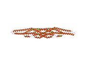 1y2o: Structure of N-terminal domain IRSp53/BAIAP2


