Dorsal root of spinal nerve
| Dorsal root of spinal nerve | |
|---|---|
 The formation of the spinal nerve from the dorsal and ventral roots | |
| Details | |
| Identifiers | |
| Latin | radix posterior nervi spinalis |
| TA98 | A14.2.00.030 |
| TA2 | 6146 |
| FMA | 5980 |
| Anatomical terminology [edit on Wikidata] | |
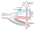
The dorsal root of spinal nerve (or posterior root of spinal nerve or sensory root)[1] is one of two "roots" which emerge from the spinal cord. It emerges directly from the spinal cord, and travels to the dorsal root ganglion. Nerve fibres with the ventral root then combine to form a spinal nerve. The dorsal root transmits sensory information, forming the afferent sensory root of a spinal nerve.
Structure
The root emerges from the posterior part of the spinal cord and travels to the dorsal root ganglion. The dorsal root ganglia contain the pseudo-unipolar cell bodies of the nerve fibres which travel from the ganglia through the root into the spinal cord.
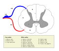
The lateral division of the dorsal root contains lightly myelinated and unmyelinated fibres of small diameter.[citation needed] These carry pain and temperature sensation. These fibers cross through the anterior white commissure to form the anterolateral system in the lateral funiculus.
The medial division of the dorsal root contains myelinated fibres of larger diameter[citation needed]. These transmit information of discriminative touch, pressure, vibration, and conscious proprioception originating from spinal levels C2 through S5. These fibers are pushed in towards the posterior median sulcus to form the gracile fasciculus and the cuneate fasciculus of the posterior column–medial lemniscus pathway. If the dorsal root of a spinal nerve were severed, it would lead to numbness in certain areas of the body.
Additional images
-
-
 Medulla spinalis
 -
 A spinal nerve with its anterior and posterior roots.
 -
 The sensory tract.
 -
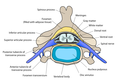
 Diagrammatic transverse section of the medulla spinalis and its membranes.
 -
 A portion of the spinal cord, showing its right lateral surface. The dura is opened and arranged to show the nerve roots.
 -
 Scheme showing structure of a typical spinal nerve.


See also
References
- ^ "Spinal Nerves - an overview". ScienceDirect Topics. Archived from the original on May 6, 2021. Retrieved 2021-05-06.
External links
- Anatomy figure: 02:04-06 at Human Anatomy Online, SUNY Downstate Medical Center - "Superior view of a section through the spinal cord within the vertebral foramen."
- Anatomy Atlases – Microscopic Anatomy, plate 06.114 - "Spinal Root Nerve Fibers"
- Dorsal root - Cell Centered Database
- v
- t
- e
| Tissue Types | |||||
|---|---|---|---|---|---|
| Cell Types |
|
| General |
|
|---|---|
| Connective tissues | |
| Neuroglia |
nerve fibers
| Parts |
| ||||||
|---|---|---|---|---|---|---|---|
| Types | |||||||
| Afferent nerve fiber/ Sensory neuron | |||||||
| Efferent nerve fiber/ Motor neuron |
| Synapse | |
|---|---|
| Sensory receptors |
| Authority control databases |
|
|---|


